
Learning Pathway
Diploma in Medical
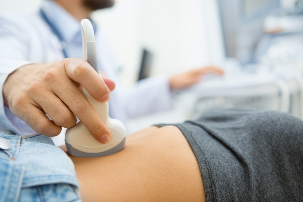
Ultrasound (DMU)
1. DMU (1 year)
Equip and empower yourself to enter into the field of medical ultrasound through the Diploma in Medical Ultrasound (DMU)
DMU course is tailored to meet the requirements of medical professionals. The course provides the students with the expertise in the imaging and confidence in interpreting & diagnosis of medical ultrasound cases in a clinical set up.


Course Speakers

Dr. Syed Muhammad Baqui Billah
MBBS, MPH, PhD Associate Professor

Dr. Md. Nazrul Islam
MBBS, DMU, MSc (MS) Course Coordinator

Dr. Mahbuba Begum
MBBS, DMU Teacher

Dr. Prianka
MBBS, DMU Teacher
Curriculum
Chapter 1| Lecture
Basics of medical ultrasound
Basic functioning of an ultrasound machine, scanning planes, cross infection and patient’s safety and documentation (image printing & report writing)
Chapter 1 | Logbook Image
2 Images from: Knobology, probe manipulation techniques, acquisition of images and orientation of image
Chapter 2 | Lecture
Ultrasound physics
Ultrasound physics: 1
Ultrasound physics: 2
Chapter 2 | Logbook Image
2 Images from: Selection of transducer, changing the frequency according to depth
Chapter 3 | Lecture
IVC ultrasound: Indication of scanning, hepatic veins and their course, Valsalva
Abdominal aorta ultrasound: Indication of scanning, aortic aneurysm, dissecting aorta
Chapter 3 | Logbook Image
2 Images from: (1) Aorta (2) IVC
Chapter 4 | Lecture
Hepatobiliary system ultrasound
Normal anatomy, developmental variants, liver parenchymal disease, solid and cystic lesions of liver, gallbladder and biliary tract diseases, diseases of spleen and pancreas
Chapter 4 | Logbook Image
4 Images from: Liver, GB, CBD
2 Image from: Pancreas
2 Images from: Spleen
Chapter 5 | Lecture
Urinary system ultrasound
Normal anatomy, developmental variants, diseases of kidney, adrenal glands, urinary bladder and prostate
Chapter 5 | Logbook Image
4 Images from: Kidney
1 Image from: Adrenal gland
1 Images from: UB
1 Images from: Prostate
Chapter 6 | Lecture
Gastrointestinal tract ultrasound
Indication of scanning, anatomy of parts of GIT, diseases of GIT
Chapter 6 | Logbook Image
2 Images from: GIT
Chapter 7 | Lecture
Pelvic organs ultrasound
Normal anatomy and related disease, developmental anomaly, diseases of uterus, ovaries and adnexa, TVS
Chapter 7 | Logbook Image
4 Images from: Uterus
4 Images from: Ovary
2 Images from: TVS
Chapter 8 | Lecture
Obstetrics: First trimester ultrasound
Indication of scanning, facts & features of a gestational sac, gestational age measuring techniques in first trimester, mean sac diameter & blighted ovum, normal & abnormal yolk sac, complications in first trimester
Chapter 8 | Logbook Image
2 Images from: GS
2 Images from: Embryo
Chapter 9 | Lecture
Obstetrics: Second & third trimester ultrasound
Estimation of gestational age in 2nd & 3rd trimester, growth scan, assessment of amniotic fluid & placenta, biophysical profile, obstetric Doppler, small for gestational age and IUGR
Chapter 9 | Logbook Image
4 Images from: BPD, FL, HC & AC
2 Images from: AFI, Placenta
2 Images from: Umbilical & MCA Doppler
Chapter 10 | Lecture
Obstetrics: Level II obstetric ultrasound (Anomaly scan)
Facts & indication, anomaly of head, anomaly of brain, anomaly of face, anomaly of neck, anomaly of spine, anomaly of chest, anomaly of heart, anomaly of abdomen, anomaly of urinary tract and skeletal
Chapter 10 | Logbook Image
8 Images from: Different parts of fetus (preferably with anomaly)
Chapter 11 | Lecture
Small part: Breast ultrasound
Application & indication of scanning, anatomy & zones of breast, lymphatic drainage of breast, advantages & disadvantages of breast ultrasound, cystic, solid & complex masses, BIRAD Scoring, reporting
Chapter 11 | Logbook Image
4 Images from: Normal breast and pathology (preferably with Doppler)
Chapter 12 | Lecture
Small part: Scrotum ultrasound
Techniques & indication of scanning, anatomy of testicle, cystic & solid masses, testicular inflammation, infarction & abscess, testicular calcifications with microlithiasis, Extra-testicular lesions, hernia, varicocele, diseases of epididymis, torsion testis, cryptorchidism, testicular trauma
Chapter 12 | Logbook Image
4 Images from: Normal testis and pathology (preferably with Doppler)
Chapter 13 | Lecture
Small part: Thyroid ultrasound
Indication of scanning, normal anatomy with sonographic findings, developmental variants, aplasia of thyroid & ectopic thyroid tissue, imaging pitfalls, nodular disease, thyroid malignancy & TIRAD score, autoimmune thyroiditis (Hashimoto thyroiditis & Graves’ disease, reporting of thyroid disease.
Chapter 13 | Logbook Image
4 Images from: Normal thyroid and pathology (preferably with Doppler)
Chapter 14 | Lecture
Vascular ultrasound
Duplex study of carotid with characterization of plaque and stenosis, Duplex study of peripheral vessels with arterial occlusive disease, DVT, varicose vein & incompetence of venous valves.
Chapter 14 | Logbook Image
Chapter 15 | Lecture
Musculoskeletal (MSK) ultrasound
Facts & indication, basics of MSK ultrasound, ultrasound of rotator cuff, ultrasound of knee joint, ultrasound of Achilles tendon, Carpal tunnel syndrome
Chapter 15 | Logbook Image
2 Images from: Tendon
2 Images from: Rotator cuff
2 Images from: Median nerve
Chapter 16 | Lecture
Lung ultrasound
Diagnostic value of different modalities in lung imaging, transducers used in lung ultrasound, patient position and scanning zones, indications, normal findings of lung ultrasound, A-line, B-line & dynamic changes during respiration, pneumothorax, pleural effusion, pneumonia, alveolar interstitial syndrome
Chapter 16 | Logbook Image
1 Image from: A-line
1 Image from: B-line
1 Image from: Pleural effusion
1 Image from: Consolidation
Chapter 17 | Lecture
Pediatric ultrasound
Developmental dysplasia of the hip (DDH), infantile hypertrophic pyloric stenosis (IHPS), neonatal brain ultrasound, intussusception
Chapter 17 | Logbook Image
2 Images from: Neonatal brain
1 Image from: Hip joint
1 Images from: Pylorus (preferably with IHPS)
Ideal for

Doctors

Practicing Sonologists
Sonographers
Pay fees

Currier Fee
For international student
International currier fee € 150
If any Bangladeshi student wants to receive his/her Diploma Certificate from the university to his/her home address, he/she will need to pay € 150 as international currier fee; e.g. via DHL, FedEx etc.
Eligibility for Admission
Bangladeshi students
- MBBS or MD
- Bangladesh Medical and Dental Council (BMDC) registration
- Available computer with internet connection
International students
- MBBS or MD or equivalent
- Registration with local authority
- Practicing as Sonographer (should have opportunity to take images to complete logbook)
- Available computer with internet connection
Note: Before enrollment please check with your local registration authority whether they will recognize this course or not.
Way to get Diploma
- Check your eligibility
- Apply for the course
- Pay fees
- Attend the lectures and complete the course
- Pass the exams
- Complete your logbook
- Pay currier fee (where applicable)